Beamline 2.4 contains two separate endstations, the NeaSpec NeaSNOM and the Agilent FTIR with focal plane array detection.
Endstation 2.4.1 NeaSpec NeaSNOM

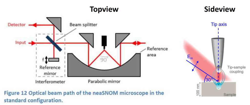
The NeaSpec endstation operates in two types of nano-resolved modes: Nano-FTIR and Nano-Imaging.
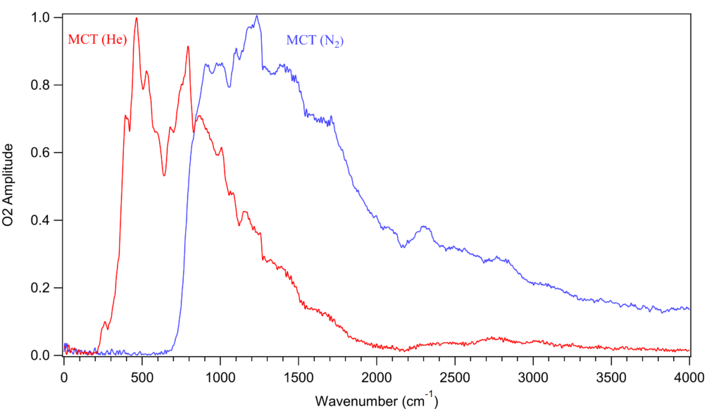
Nano-FTIR or SINS utilizes the ultrabroadband synchrotron IR light to collect spectra with spatial resolution defined by the width of the AFM probe (25 nm is typical). The instrument can be operated remotely. We typically use two different detectors. A liquid helium-cooled MCT detector for frequencies between ~200 and 2000 cm-1 and a liquid nitrogen-cooled MCT detector for frequencies between 750 and 4000 cm-1.


Nano-Imaging is a complementary technique which uses laser-based light sources for mapping the electric field or specific absorption resonances on the topography. Nano-imaging is available to users when the synchrotron is not operating. We presently maintain the following light sources for Nano-Imaging:
| Laser | Wavenumbers (cm-1) | μm | meV |
| CO2 | 900 – 1000 | 10 – 11 | 113 – 124 |
| QCL | 1130 – 1938 | 5.2 – 8.8 | 140 – 240 |
For more information on the terminology used here, please see the FAQ page.

The user guide for the Nano-FTIR instrument can be found on the Manuals and Software page.
Coming Soon:
Kelvin Force Probe Microscopy (KPFM) to resolve variations in workfunction over a surface
Piezo Force Microscopy (PFM) to image grain boundaries and domains in piezoelectric materials.

Agilent FTIR
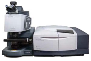
The Agilent FTIR is not presently connected to the synchrotron; instead, an internal globar source is used. This system is available to users by applying for access to Beamline 2.4 and specifying this endstation.
The 128 x 128 pixel focal plane array detector (FPA) enables rapid hyperspectral imaging.
| Objective | NA | High Mag | Field of View | Effective Pixel Size |
|
15x |
0.62 | No | 704 μm x 704 μm | 5.5 μm |
| 15x | 0.62 | Yes | 141 μm x 141 μm | 1.1 μm |
| 25x | 0.81 | No | 420 μm x 420 μm | 3.3 μm |
| 25x | 0.81 | Yes | 85 μm x 85 μm | 0.66 μm |
The user guide for this microscope can be found here.