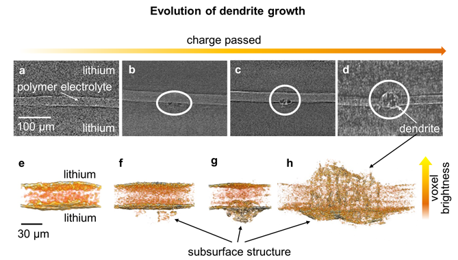
Lithium-ion batteries, popular in today’s electronic devices and electric vehicles, could gain significant energy density if their graphite anodes were replaced with lithium metal anodes. But there’s a major concern with substituting lithium—when the battery cycles, microscopic fibers of the lithium anodes (“dendrites”) form on the surface of the lithium electrode and spread across the electrolyte until they reach the other electrode. An electrical current passing through these dendrites can short-circuit the battery, causing it to overheat or catch fire. The dendrites are one of the major reasons for lithium failure, but they are very difficult to see. This is one of the reason why efforts to curtail dendrite growth have thus far met with little success. But researchers have recently discovered that the x-ray microtomography capabilities at ALS Beamline 8.3.2 can give them a novel view of dendrite growth that’s likely to provide the insight needed to stop it.

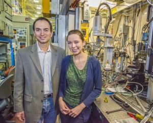
The ALS researchers, led by Nitash Balsara, a faculty scientist with Berkeley Lab’s Material Sciences Division and a professor of chemical engineering at UC Berkeley, used the microtomography technique to gather data about the lithium batteries throughout their various stages of cycling. What they discovered was that during the early stages of development, the bulk of dendrite material lies below the surface of the lithium electrode, underneath the electrode/electrolyte interface. Using the x-ray microtomography technique, the researchers observed the seeds of dendrites forming in lithium anodes and growing out into a polymer electrolyte during cycling. It was not until the advanced stages of development that the bulk of dendrite material was in the electrolyte. Balsara and his colleagues suspect that non-conductive contaminants in the lithium anode trigger dendrite nucleation.
The tremendous energy density of lithium and the metal’s remarkable ability to move lithium ions and electrons in and out of an electrode as it cycles through charge/discharge make it an ideal anode material. Until now, researchers have studied the dendrite problem using various forms of electron microscopy. This is the first study to employ microtomography using monochromatic beams of high energy or “hard” x-rays, ranging from 22 to 25 keV, at ALS Beamline 8.3.2. This technique allows non-destructive three-dimensional imaging of solid objects at a resolution of approximately one micron.
“The capabilities at the ALS allowed us to see into the battery without destroying it, which was key to extracting this data,” says Katherine Harry, a graduate student in Balsara’s group at UC Berkeley. “We were able to watch how the dendrites form starting inside the electrodes themselves, which you can’t do with other techniques because lithium metal is opaque to electrons but transparent to x-rays.”
Harry explains that the ALS technique is particularly useful for industry because it allows researchers to watch how dendrites form and grow under a variety of conditions. Indeed, the technique Harry used has already attracted new industrial users to Beamline 8.3.2.
“You can see how factors such as temperature affect dendrite growth and you can quantify that,” she says. “Once you really understand the relationship between the various parameters and dendrites, you can control dendrite growth.”

The success of the technique for this particular type of battery research was enabled by the unique capabilities of Beamline 8.3.2. “There are a lot of lab-based (non-synchrotron) tomography systems,” explains ALS beamline scientist Dula Parkinson. “But one of the things that’s special about the ALS is the high x-ray flux that allows us to use a monochromator to select a single x-ray energy for an experiment.”
Using the high-energy x-rays from most lab-based tomography systems, materials like carbon and lithium are nearly invisible. On the other hand, Beamline 8.3.2 can select an x-ray energy with more or less penetrating power. In this experiment, an x-ray energy was chosen that was high enough to penetrated the battery and its packaging, but still low enough to yield good contrast for the components of interest.
Harry and fellow researchers are continuing their dendrite research at the ALS with a focus on in-situ experiments, trying to understand the relationship between a variety of parameters and the morphology of dendrite growth. She says her time at the ALS and the assistance of beamline scientists Parkinson and Alastair MacDowell have been invaluable to her research: “The ALS beamline scientists are experts and so helpful and helped me get results so much faster.”
Balsara is the corresponding author of a paper describing this research in Nature Materials titled “Detection of subsurface structures underneath dendrites formed on cycled lithium metal electrodes.” Co-authors are Katherine Harry, Daniel Hallinan, Dilworth Parkinson, and Alastair MacDowell.