Advanced ceramic composites can withstand the ultrahigh operational temperatures projected for hypersonic jet and next-generation gas-turbine engines, but real-time analysis of the mechanical properties of these space-age materials at ultrahigh temperatures has been a challenge—until now. Researchers have developed the first testing facility that enables microtomography of ceramic composites under controlled loads at ultrahigh temperatures and in real-time. Using this facility, they have fully resolved sequences of microcrack damage as cracks grow under load at temperatures several hundred degrees higher than previously possible. The observations are key ingredients of the high-fidelity simulations used to compute failure risks under extreme operating conditions.
Ceramics made from clay have been used as construction materials for thousands of years and are renowned for their ability to resist damage from water, chemicals, oxidation, and—most importantly—heat. Ceramics can stand up to temperatures that would melt most metals. However, traditional ceramics also suffer from a serious deficit—brittleness. Today’s advanced ceramics for extreme structural applications are much stronger and tougher. They’re reinforced with ceramic fibers to form composites that can be structured along the lines of natural materials such as teeth and bone. Jet or gas-turbine engines made from ceramic composites would weigh considerably less than today’s engines and operate at much higher temperatures. This translates into far greater fuel efficiencies and reduced pollution.
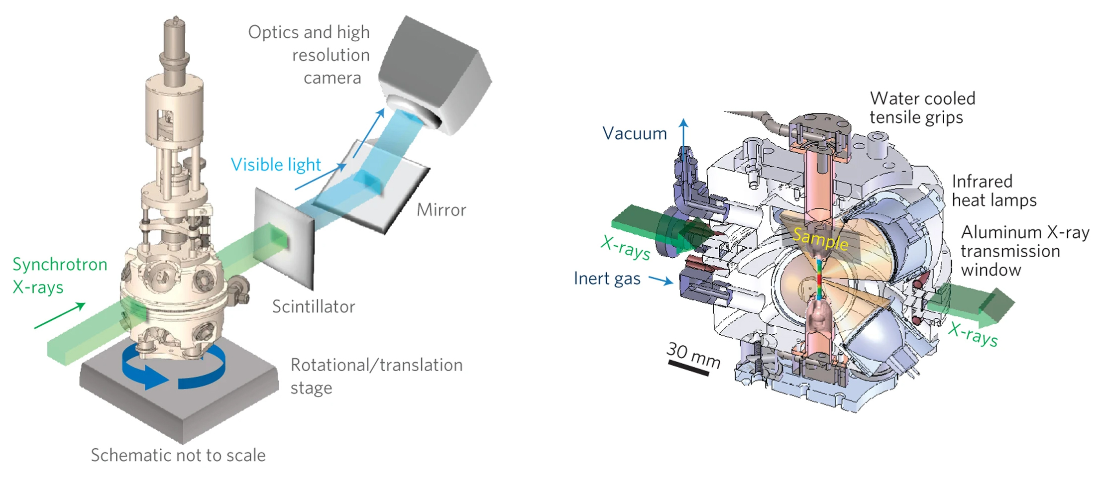
Silicon carbide ceramics, reinforced with woven ceramic fibers, can improve jet-engine efficiency by tolerating higher operating temperatures. But first, the material must be thoroughly tested and its behavior understood at the microscopic level. This video describes a newly developed high-temperature microtomography cell that allows scientists to study these materials in 3D, at micron scale, under load, and at high temperatures.
Still, while ceramic composites are far less prone to fracture than their clay ancestors, tiny cracks can form and grow within their complex microstructures, creating potentially catastrophic problems. Like teeth and bone, ceramic composites achieve robustness through complexity, with their hierarchical, hybrid microstructures impeding the growth of local damage and preventing the large fatal cracks that are characteristic of brittle materials. However, complexity in composition brings complexity in safe use. For ceramic composites in ultrahigh-temperature applications, especially where corrosive species in the environment must be kept out of the material, relatively small cracks, on the order of a single micron, can be unacceptable.
Exactly how microcracks are restrained by the tailored microstructures of a ceramic composite is the central question for the materials scientist seeking the optimal composition or architecture, and the design engineer who must predict the failure envelope. The only reliable way to answer this question is through measurements made at ultrahigh temperatures.
ALS Beamline 8.3.2, which is powered by a 6-tesla superbend magnet, is designed for x-ray computed microtomography, a technology that provides nondestructive 3D imaging of solid objects at a resolution of approximately one micron. The research team designed and placed into this beamline a unique tensile-testing rig that can pull or compress a small, cylindrical sample in ultrahigh-temperature environments (up to 1750°C). The rig can also operate in vacuum or in atmospheres that are inert or oxidizing. They then obtained 3D tomographic images of samples consisting of a silicon carbide ceramic matrix, reinforced with silicon carbide fibers, at sufficient resolution to observe the real-time formation and growth of microcracks and other forms of internal damage as a function of load.
The data obtained contain complete quantitative information on crack paths, crack surface areas and orientations, spatial variations in the crack-opening displacements, statistics of relative spatial location of cracks, and microstructural heterogeneities within the sample volume. All these parameters are critical in any analysis of fracture as they govern the toughness of the material.
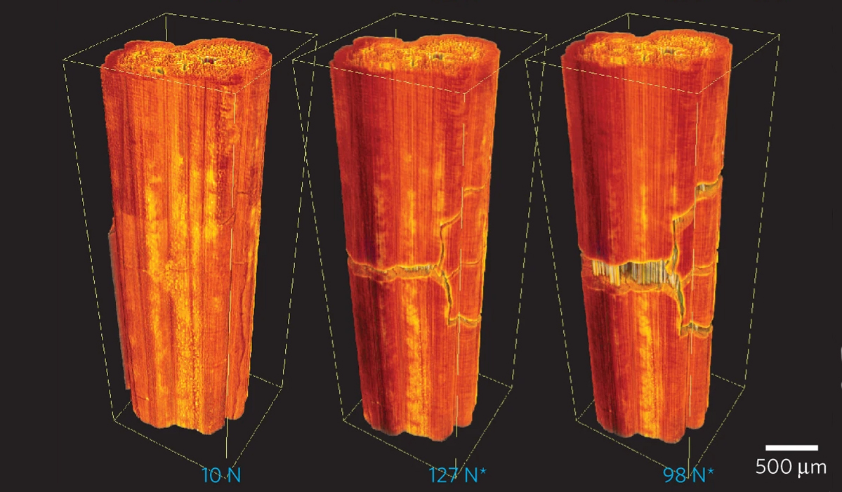
Three-dimensional volume-rendered microtomography images from a specimen tested at 1750°C at several applied loads
(10, 127, and 98 newtons, respectively). *Load reading after first matrix crack initiated.
The results provide vital information about the underlying failure mechanisms in ceramic composites that can be used to optimize their performance and predict structural integrity and safe lifetime. The capacity for validating virtual testing models through direct, real-time, noninvasive experimental observations should greatly advance our understanding and help promote the technological innovation of ceramic composites.
Contact: Rob Ritchie
Researchers: H.A. Bale (Univ. of California, Berkeley); A. Haboub, A.A. MacDowell, J.R. Nasiatka, and D.Y. Parkinson (ALS); B.N. Cox and D.B. Marshall (Teledyne Scientific Company); and R.O. Ritchie (Univ. of California, Berkeley, and Berkeley Lab).
Funding: Air Force Office of Scientific Research, National Aeronautics and Space Administration, and Teledyne Scientific Company. Operation of the ALS is supported by the U.S. Department of Energy, Office of Basic Energy Sciences.
Publication: H.A. Bale, A. Haboub, A.A. MacDowell, J.R. Nasiatka, D.Y. Parkinson, B.N. Cox, D.B. Marshall, and R.O. Ritchie, “Real-time quantitative imaging of failure events in materials under load at temperatures above 1,600 °C,” Nat. Mater. 12, 40 (2013).
ALS SCIENCE HIGHLIGHT #268
![]()
![]()