A hallmark of Alzheimer’s disease, the most prevalent type of dementia, is the formation of insoluble deposits in the brain. The deposits, or plaques, consist of aggregated amyloid-β peptides that may impair function. Alongside plaque formation, disrupted metal homeostasis increases iron levels in several regions of Alzheimer’s brains; high concentrations of chemically reduced forms of iron are associated with pathological features of the disease.
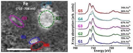
Iron in a healthy living brain is primarily stored in a way that minimizes the production of free radicals. Researchers suspected that chemical reduction of this iron releases free radicals, causing the damage observed in Alzheimer’s disease. In this work, the researchers extracted amyloid plaques from the donated brains of two deceased individuals who had Alzheimer’s, then performed synchrotron scanning transmission x-ray microscopy (STXM) at ALS Beamline 11.0.2 and Diamond Light Source to locate and characterize nanoscale inclusions of iron oxide in the samples in unprecedented detail.
The STXM analysis revealed ferrous iron, mixed-valence iron oxide magnetite, and even zero-valent iron, indicating that excessive chemical reduction of iron occurred at the amyloid plaque sites. Ptychography at the ALS provided unique insight into the morphology of a magnetite/maghemite inclusion, indicating a faceted structure consistent with a biological (rather than industrial) origin. Multiple phases of calcium were also observed in the plaques, raising questions about whether they are associated with the disrupted neuronal calcium regulation and signaling that occurs in Alzheimer’s disease. These findings closely link iron reduction to amyloid plaque formation and raise challenging questions about the contribution of free radicals from disrupted metal homeostasis in Alzheimer’s disease.

J. Everett, J. F. Collingwood, V. Tjendana-Tjhin, J. Brooks, F. Lermyte, G. Plascencia-Villa, I. Hands-Portman, J. Dobson, G. Perry, and N. D. Telling, “Nanoscale synchrotron X-ray speciation of iron and calcium compounds in amyloid plaque cores from Alzheimer’s disease subjects,” Nanoscale 10 (2018), doi:10.1039/c7nr06794a.