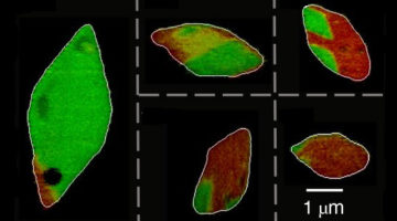
Experiments revealed that lithium ions unexpectedly flow along the surfaces of electrode particles, boosting the growth of lithium “hot spots” that shorten battery life. The results correct decades’ worth of assumptions and will help improve battery design, potentially leading to a new generation of lithium-ion batteries. Read more »![]()
ALS Work Using Microscopy/Imaging
These techniques use the light-source beam to obtain pictures with fine spatial resolution of the samples under study and are used in diverse research areas such as cell biology, lithography, infrared microscopy, radiology, and x-ray tomography.
Comparative morphology of cheliceral muscles using high‐resolution X‐ray microcomputed‐tomography in palpimanoid spiders

Spiders are important predators in terrestrial ecosystems, yet we know very little about their principal feeding structures—the chelicerae—an extremely important aspect of spider biology. Here, using micro‐Computed‐Tomography scanning techniques, researchers perform a comparative study to examine cheliceral muscle morphology in six different spider specimens. Read more »
Memristor Collaboration between ALS and Hewlett Packard Labs Propels Theory to Application
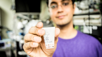
The development of an idea into a commercial product can take decades, a timeframe that allows contributions from multiple generations of scientists and requires investment in basic research. Collaborative research between the ALS and Hewlett Packard Labs has advanced the memristor, a device that can store information using little to no power. Read more »
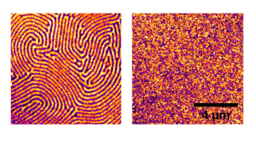
Magnetic Skyrmions: Current‐Induced Skyrmion Generation through Morphological Thermal Transitions in Chiral Ferromagnetic Heterostructures

Magnetic skyrmions are particle‐like chiral twists of the magnetization that promise advances in spin‐based data storage and logic device applications. In this article, researchers examine current‐induced generation of skyrmions in heavy‐metal/ferromagnet multilayers and show that Joule heat pulses can drive topological transitions in magnetic textures and enable skyrmion creation on nanosecond timescales. Read more »
Tunable Ferromagnetism in a 2D Material at Room Temperature
Researchers combined soft x-ray spectroscopy and microscopy to demonstrate the tunable ferromagnetic characteristics of a two-dimensional layered material at room temperature. The results open up exciting opportunities for the use of such materials in low-power spintronics, high-density magnetic storage, and flexible electronics. Read more »
Expanding the Infrared Nanospectroscopy Window
An innovative infrared-light probe with nanoscale spatial resolution has been expanded to cover previously inaccessible far-infrared wavelengths. The ability to investigate heterogeneous materials at nanometer scales and far-infrared energies will benefit a wide range of fields, from condensed matter physics to biology. Read more »![]()
![]()
Near-field infrared nanospectroscopy and super-resolution fluorescence microscopy enable complementary nanoscale analyses of lymphocyte nuclei
Recent super-resolution fluorescence microscopy studies have revealed significantly altered nuclear organization between normal lymphocyte nuclei and those of classical Hodgkin’s lymphoma. Reported here are the first near-field IR imaging of lymphocyte nuclei, and far-field IR imaging results of whole lymphocytes and nuclei from normal human blood. Read more »
Miscibility–Function Relations in Organic Solar Cells: Significance of Optimal Miscibility in Relation to Percolation

In this article, Ye et al. present the determination of liquidus miscibility and its temperature dependence of organic films by scanning transmission x‐ray microscopy and outline an approach to convert liquidus miscibility to an effective Flory‐Huggins interaction parameter χ, which will pave a way to predict morphology and processing strategies of polymer solar cells. Read more »
Nanoscale Characterization of Iron and Calcium in the Alzheimer’s Brain
The amyloid plaques that accumulate in Alzheimer’s disease contain stores of iron. Using scanning transmission x-ray microscopy, researchers have characterized the iron’s chemical states in unprecedented detail. Their nanoscale analysis revealed excessive chemical reduction of the iron, which can release damaging free radicals. Read more »
Infrared Beams Show Cell Types in a Different Light

By shining highly focused infrared light on living cells, scientists hope to unmask individual cell identities and to diagnose whether the cells are diseased or healthy. Their focus is on developing a rapid, noninvasive way to easily identify cell types and features within living cells, to aid in biological and medical research. Read more »
- « Previous Page
- 1
- …
- 9
- 10
- 11
- 12
- 13
- …
- 17
- Next Page »