<<Return to 2025 User Meeting Agenda
 |
Greg HuraScience Deputy, Molecular Biology Biosciences Division, LBNL Probing Complex Chemistry at the Molecular Level with Vibrational Spectroscopy and X-Ray Tools Bio Greg Hura is the Science Deputy of the Molecular Biophysics and Integrated Bioimaging Division at Lawrence Berkeley National Laboratory. Hura’s research focuses on understanding the structural mechanisms of biological macromolecular machines and their implications at larger length scales or for novel design. At the Advanced Light Source (ALS), he is part of the Structurally Integrated Biology for the Life Sciences (SIBYLS) group, which operates Beamline 12.3.1. He also serves as the Biosciences Thrust Area Representative and is the local lead of the Integrated Diffraction Analysis Technologies program. Hura is a co-principal investigator in the NIH-funded ALS-ENABLE program and leads the BRaVE Taskforce 5 effort. He has been at the Lab since 1996 and is an adjunct professor of chemistry and biochemistry at UC Santa Cruz. |
 |
Guiliang XuChemist Synchrotron Characterization of Lithium-Ion Batteries and Beyond: From Material Discovery to Cell Diagnostics Bio Guiliang Xu is a chemist in the Chemical Sciences and Engineering Division at Argonne National Laboratory and a CASE scientist at the Pritzker School of Molecular Engineering at the University of Chicago. He received his Bachelor’s degree in 2009 and Ph.D. in 2014 from Xiamen University. Dr. Xu has over 15 years of research experience in the design, synthesis and processing of multifunctional materials for advanced energy storage systems, including lithium-ion, sodium-ion, lithium-sulfur, and solid-state batteries. He utilizes cutting-edge characterization techniques to elucidate the fundamental relationships between material structure and electrochemical performance, enabling the development of safer, higher-energy-density battery materials. |
 |
Andrzej JoachimiakArgonne Distinguished Fellow New Structural Biology Abstract Breakthroughs in genome sequencing and structural biology are revolutionizing our understanding of complex biological systems and bioenvironments. By decoding DNA, RNA, and protein sequences, scientists can gain insights into how living organisms function. However, to fully grasp these systems, it’s crucial to study the structures of proteins and other biological molecules. Advancements in technology, such as new X-ray synchrotron facilities, have greatly enhanced our ability to visualize protein structures. These facilities use powerful beams to reveal the detailed architecture of proteins, which is essential for understanding their roles in biological processes. Global efforts in Structural Genomics are streamlining the process of determining protein structures. These initiatives use cost-effective techniques in bioinformatics and molecular biology to efficiently map out protein structures. Cryo-electron microscopy (Cryo-EM) is another cutting-edge tool that allows scientists to study large and complex molecular assemblies. Solution studies and molecular dynamic simulations expand our understanding of assembly and regulation. Moreover, public databases filled with protein structures are fueling the development of sophisticated algorithms like AlphaFold and Rosetta. These algorithms can predict protein structures from scratch, complementing experimental methods and accelerating discoveries. The shared knowledge from these efforts not only helps us understand how biological systems evolve but also aids in designing new proteins and synthetic biological functions. As technology continues to advance, particularly in areas like supercomputing and artificial intelligence, our understanding of biology is poised to reach new heights. Despite ongoing challenges, the future of genomics, structural biology and artificial intelligence holds immense promise for scientific discovery and innovation. Bio Dr. Andrzej Joachimiak is an expert in synchrotron-based X-ray crystallography and structural biology. At Argonne, he has made significant contributions to the high-throughput crystallography using synchrotron radiation and the development of state-of-the-art facilities for macromolecular crystallography. For the past 30 years, he served as the Director of Structural Biology Center, one of the world’s leading facilities for protein structure determination. In the past 24 years, the NIH has supported his research in this area as a part of the Protein Structure Initiative, the Regional Center of Excellence, and the Structural Genomics/Biology of Infectious Diseases programs. He has been involved in several large and highly successful collaborative efforts to study proteins, enzymes and drug discovery relevant to human diseases, including cancer and more recently SARS-CoV-2. His laboratory contributed many high-resolution crystal structures of viral proteins and complexes with interacting partners, antibodies and small ligands. He directed the Argonne-based Midwest Center for Structural Genomics (MCSG), a highly successful program and major component of NIH funded Protein Structure Initiative that developed methods in protein expression and purification, crystallization and data collection using synchrotron radiation. In addition to his duties at Argonne, Andrzej is also a Professor and Senior Scientist of the Consortium for Advanced Science and Engineering at the University of Chicago, Adjunct Professor and Senior Fellow at the Institute of Science and Engineering at Northwestern University, CoPI, DOE Biopreparedness Research Virtual Environment, Taskforce 5. |
 |
Yury GogotsiDistinguished University Professor and Chair in the Department of Materials Science and Engineering Challenges and Opportunities in Characterization of MXenes Abstract MXenes are a large and quickly growing family of 2D materials with over 100 compositions reported, not counting diverse surface terminations [1]. There are tens of thousands of research papers and dozens of books originating from over 100 countries; about 10,000 patents were filed in the past decade (in fact, the majority just in the past 5-6 years) [2]. While for many materials that could indicate a mature state of research and even saturation of the field, in the case of MXenes, this is just the beginning. We are only scratching the very surface of this emerging field. The number of possible MXene compositions is infinite if one considers solid solutions on M and X sites, high-entropy MXenes, and combinations of surface terminations. New subfamilies of in- and out-of-plane ordered MXenes, oxycarbides, 2D borides, and silicides further expand the family of non-oxide 2D materials based on transition metals. MXenes have led us into the world of atomistically designed materials. This variety of structures and compositions creates challenges in characterization. It is often necessary to use multiple advanced characterization techniques within a single study, and each characterization technique has its own quirks, pitfalls, and benefits when applied to MXenes [3]. This presentation focuses on the utilization of X-ray PDF, powder diffraction, X-ray photoelectron spectroscopy, Raman and IR spectroscopy, electron microscopy/spectroscopy, and a number of other techniques to understand MXenes and determine their composition, structure, and properties. Bio Yury Gogotsi is a Distinguished University Professor and the Charles T. and Ruth M. Bach Endowed Chair in the Department of Materials Science and Engineering. He also serves as Director of the A.J. Drexel Nanomaterials Institute. He received his MS (1984) and PhD (1986) from Kyiv Polytechnic and a DSc degree from the National Academy of Sciences of Ukraine (1995). Together with his students and colleagues, he has made principal contributions to the development of materials for electrochemical capacitors and other energy storage devices, discovered MXenes, demonstrated the tuning of structure and porosity of carbide-derived carbons, and developed new processes for the synthesis, surface modification, and purification of nanotubes and nanodiamonds. He also published the first microscopic observation of water inside carbon nanotubes, discovered polygonal nanotubes (graphite polyhedral crystals), and shaped the field of high-pressure surface science. He is recognized as a Highly Cited Researcher in Materials Science and Chemistry and a Citations Laureate in Physics by Clarivate. He has received numerous awards for his research, including the Blaise Pascal Medal from the European Academy of Sciences, the Ceramic Prize from the World Academy of Ceramics, the Materials Research Society (MRS) Medal, the American Chemical Society (ACS) Award in the Chemistry of Materials, etc. He has been elected a Fellow of the National Academy of Inventors, the World Academy of Ceramics, the European Academy of Sciences, Academia Europaea, and many professional societies. He holds honorary doctorates from several European Universities. |
 |
Tanja BosakProfessor of Geobiology in the Department of Earth, Atmospheric and Planetary Science Evaluating Microbial Imprints on Minerals in Carbonate Deposits from Ancient and Modern Earth Abstract Some of the oldest records of life and habitable environments on Earth contain carbonate minerals. To understand how early microbes lived and interacted with their surroundings, we study modern microbial processes that shape carbonate grains and structures or enrich trace or even major elements and compare them to the signals preserved in carbonate deposits that are more than two billion years old. Carbonaceous globules within laminated and lithified carbonate rocks from the 2.7-billion-year-old Tumbiana Formation in Western Australia that are enriched in arsenic were interpreted as remnants of early arsenic-metabolizing microbes that built these carbonate rocks. These arsenic-enriched globules inspire the following two hypotheses: 1. Arsenic-metabolizing microbes bound arsenic to extracellular microbial polymers while alive or 2. Hydrothermal fluids delivered arsenic to the carbonate-bound organic matter after the cessation of microbial metabolic activity. Experiments with modern microbes reveal that the dead organic matter exposed to hot, As-containing fluids has a much higher affinity for arsenic than living microbial mats or carbonate minerals. Mapping of arsenic and other elements by laser ablation- inductively coupled mass spectrometry and synchrotron XRF and measurements of arsenic speciation in the rock samples by K-edge XANES reveal the general dearth of arsenic in all but one locality from the Tumbiana Formation and the association of the detectable arsenic with sulfide minerals. These experiments and analyses favor the interpretation of previously detected arsenic enrichments as products of arsenic delivery by hydrothermal fluids, rather than remnants of microbial metabolisms that evolved by 2.7 billion years ago. Bio Tanja Bosak is professor of geobiology in the Department of Earth, Atmospheric and Planetary Sciences at MIT. Bosak’s work in experimental geobiology asks how microbial processes leave chemical, mineral, and morphological signals in sedimentary rocks. The research in her lab combines microbiology, materials science and sedimentology to explore modern biogeochemical and sedimentological processes, interpret the coevolution of life and the environment during the first 80% of Earth’s history, and search for signs of past life or prebiotic processes on Mars. Bosak’s awards include the 2007 Subaru Outstanding Woman in Science Award from the Geological Society of America and the 2011 James B. Macelwane Medal from the American Geophysical Union. Bosak is an American Geophysical Union Fellow and a member of NASA’s Mars 2020 Project Science Group. Born in Croatia, Bosak earned a BS in geophysics from Zagreb University and a PhD in geobiology from the California Institute of Technology. Webpage: Bosak Lab |
Shirley Award Speakers
The X-Ray Files: Solving Science Cold Cases with X-Ray Diffraction
 |
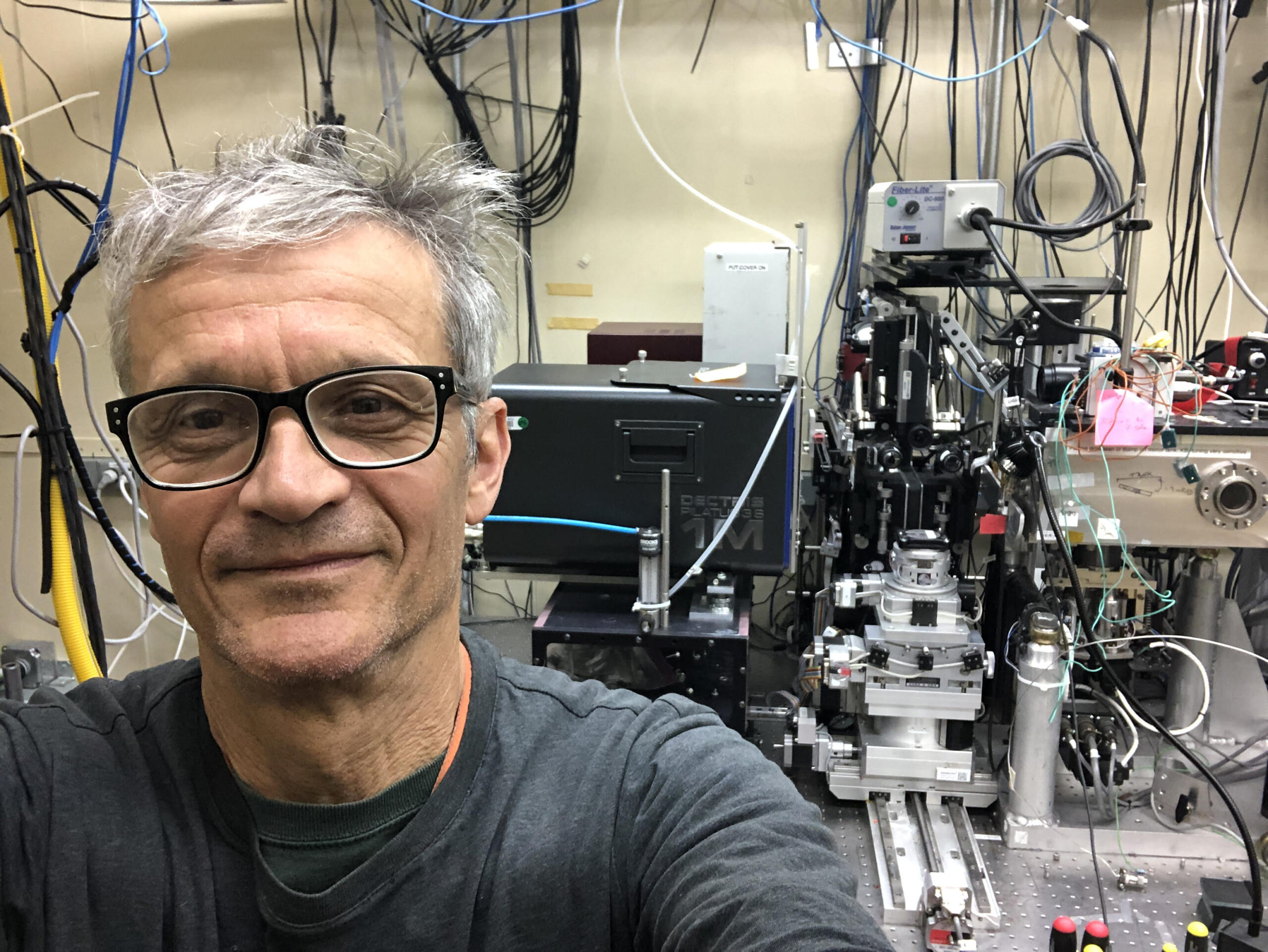
Nobumichi TamuraSenior Scientist Bio Nobumichi Tamura is a Senior Scientist in the Diffraction and Imaging Program in the Advanced Light Source Division at Lawrence Berkeley National Lab. Tamura received his Ph.D. in Materials Science from the Institut Polytechnique de Grenoble in 1993. After working for one year at Oak Ridge National Laboratory, he joined the Advanced Light Source (ALS) at Lawrence Berkeley National Laboratory, where he has been a staff scientist in the Photon Science Diffraction Group for over 25 years. His research focuses on synchrotron-based X-ray Laue microdiffraction techniques for investigating the structural properties of crystalline materials. He specializes in high-resolution mapping of strain, stress, and crystallographic orientation in polycrystalline and single-crystal samples. His work spans a broad range of materials, including metals, semiconductors, ceramics, and geological specimens. Tamura has played a key role in developing experimental methods and analytical tools, such as the X-ray Microdiffraction Analysis Software (XMAS), for interpreting complex Laue diffraction patterns. His techniques are widely used to study deformation mechanisms, grain boundary behavior, and defect structures at the microscale. Through these efforts, he has contributed to advancing the understanding of material behavior under mechanical and thermal stress. In 2019, the ALS User Executive Committee recognized Tamura with the Klaus Halbach Award for “the development of software for analysis of microdiffraction data,” critical to global science programs. |
 |
Martin KunzSenior Scientist Bio Dr. Martin Kunz is a Senior Scientist in the in the Diffraction and Imaging Program in the Advanced Light Source Division at Lawrence Berkeley National Lab. Kunz serves as beamline scientist on Beamline 12.2.2, specializing in non-ambient condition x-ray diffraction. With over two decades of experience in high-pressure research, Kunz focuses on crystal chemistry, mineral physics, and structure-property relationships in Earth materials. Kunz earned his Ph.D. in Earth Science from the University of Bern, Switzerland in 1991, and has built an international career spanning institutions in Switzerland, Canada, France, and the United States. His expertise in high-pressure mineralogy has been recognized with prestigious awards including the Paul Niggli-Medal (1995), the European Union of Mineralogy Medal for excellence in research (2004), and Fellowship in the Mineralogical Society of America (2005). Kunz’s current work involves developing and applying advanced x-ray diffraction techniques to study materials relevant to Earth’s interior, including crystal Chemistry, mineral Physics, rock weathering, and structural properties in Earth materials. |
 |
Simon TeatStaff Scientist Bio Simon Teat is a Staff Scientist in the Diffraction and Imaging Program in the Advanced Light Source (ALS) Division at Lawrence Berkeley National Lab and is the beamline scientist for 12.2.1, specializing in Chemical Crystallography. Since joining Berkeley Lab in 2006, Teat has been instrumental in advancing synchrotron-based chemical crystallography techniques and establishing world-class capabilities for small-molecule structure determination. His research focuses on the development of techniques in synchrotron chemical crystallography, with particular expertise in crystallography of the actinides, single crystal gas absorption studies, and the structure of magnetic coordination compounds. Teat’s work spans the use of anomalous scatter in materials chemistry, variable temperature studies, and phase transitions, making him a leading figure in applying advanced x-ray techniques to challenging chemical problems. |
<<Return to 2025 User Meeting