SCIENTIFIC ACHIEVEMENT
Researchers discovered that, in the nanoscale structure of human enamel (the hard outer layer of teeth), slight crystal misorientations serve as a natural toughening mechanism.
SIGNIFICANCE AND IMPACT
The results, obtained for the most part at the Advanced Light Source (ALS), help explain how human enamel can last a lifetime and provides insight into strategies for designing similarly tough bio-inspired synthetic materials.

A question to chew on
The enamel that covers the exposed surface of human teeth is the hardest tissue in the human body. Incredibly, this protective layer enables our teeth to last a lifetime. In contrast, mouse teeth grow continuously as they get worn down. In sharks and parrotfish, a new row of teeth is always ready to move forward and take over for failing ones. In crocodiles, when a tooth falls out, another one erupts. None of this happens with permanent teeth in humans, despite the fact that our teeth must withstand chewing pressures on the order of 1 GPa—comparable to pressures found 30 km underground—applied hundreds of times each day for a lifetime. How does enamel achieve such spectacular performance, despite the tremendous pressures to which it is exposed?
The hidden structure of enamel
Human tooth enamel is a hierarchical material with an intricate organization that is key to its mechanical performance. It is composed of hydroxyapatite (HAP), a biomineral of calcium and phosphate that forms long, thin nanocrystals about 50 nm wide and many microns long. In human enamel, these nanocrystals are bundled together into rods about 5 microns in diameter, with the long axes of the nanocrystals aligned with that of the rods.
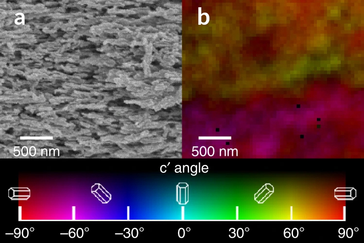
Because the parallel alignment of the nanocrystals in the rods is easy to see using scanning electron microscopy (SEM), many experts had assumed that the alignment extended to the HAP crystals’ c-axes—a well-defined direction in hexagonal HAP crystals. In fact, data about the c-axis orientations, obtained using linearly polarized x-rays, show that this is not the case.
PIC mapping the angles

In this work, researchers studied the enamel from samples of third molars (“wisdom teeth”) extracted from young adults. At ALS photoemission electron microscopy (PEEM) Beamline 11.0.1.1, variable linearly polarized light was used to create polarization-dependent imaging contrast (PIC) maps that use color to reveal variations in the orientations of the HAP crystals’ c-axes. While PIC mapping has been used extensively for carbonate biominerals, it has only recently started to be used for a calcium phosphate such as HAP. Extensive spectroscopy and theoretical modeling was done to understand the best source of dichroism contrast in HAP and its molecular-scale origins. The resulting PIC maps showed that, while the HAP nanocrystals are indeed morphologically parallel, the c-axes of adjacent nanocrystals were most frequently misoriented by 1° to 30°, with an overall variation within each rod of 30° to 90°.
Stopping cracks in their tracks
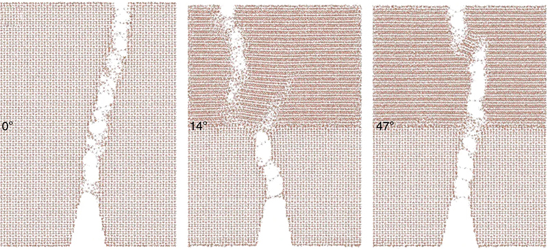
To better understand the possible function of this misorientation, the researchers performed molecular dynamics simulations to see if misoriented crystals deflect cracks at interfaces. Surprisingly, the results showed that, not only do misoriented crystals deflect cracks, but small misorientation angles are better at it than larger angles. The slight misorientations serve as an effective natural crack-deflecting mechanism, confining damage to the nanoscale and preventing propagation to the micro- and millimeter scales, thus protecting the enamel material from catastrophic failure. Such detailed insights into the structural relationships in enamel at multiple scales are extremely important for our understanding of enamel function and for the development of bio-inspired materials with greater fracture resistance.


Contact: Pupa Gilbert
Researchers: E. Beniash (University of Pittsburgh); C.A. Stifler, C.-Y. Sun, and P.U.P.A. Gilbert (University of Wisconsin–Madison); and G.S. Jung, Z. Qin, and M.J. Buehler (Massachusetts Institute of Technology).
Funding: U.S. Department of Energy, Office of Science, Basic Energy Sciences Program (DOE BES); National Science Foundation, National Institutes of Health, Office of Naval Research, and Air Force Office of Scientific Research. Operation of the ALS is supported by DOE BES.
Publication: E. Beniash, C.A. Stifler, C.-Y. Sun, G.S. Jung, Z. Qin, M.J. Buehler, and P.U.P.A. Gilbert, “The hidden structure of human enamel,” Nat. Commun. 10, 4383 (2019), doi:10.1038/s41467-019-12185-7.
ALS SCIENCE HIGHLIGHT #409