X-ray microscopy is powerful in that it can probe large volumes of material at high spatial resolution with exquisite chemical, electronic, and bond orientation contrast. The development of diffraction-based methods such as ptychography has, in principle, removed the resolution limit imposed by the characteristics of the x-ray optics. Using soft x-ray ptychography, researchers at the ALS have demonstrated the highest-resolution x-ray microscopy ever achieved by imaging five-nanometer structures.

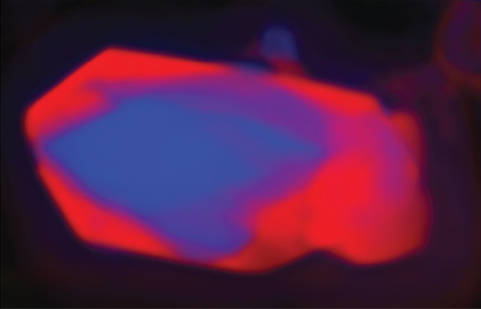
At the ALS, a collaboration of researchers used ptychographic imaging to map the chemical composition of lithium iron phosphate nanocrystals after partial delithiation. The results yielded important new insights into a material of high interest for electrochemical energy storage. Lithium iron phosphate is widely studied for its use as a cathode material in rechargeable lithium-ion batteries. In using ptychographic techniques to map the chemical composition of lithium iron phosphate crystals, ALS researchers found a strong correlation between structural defects and chemical phase propagation.

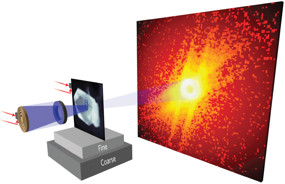
In ptychography, a combination of multiple coherent diffraction measurements is used to obtain 2D or 3D maps of micron-sized objects with high resolution and sensitivity. Because of the sensitivity of soft x-rays to electronic states, ptychography can be used to image chemical phase transformations and the mechanical consequences of those transformations that a material undergoes.
Ptychography uses coherent diffraction patterns recorded from fields of illumination that are physically overlapped on an extended sample, with known separation. This information is used in the iterative reconstruction of phase, and results in a robust and unique solution that gives the local complex refractive index of the object and the complex illumination function. Through phase retrieval from diffraction data, ptychography can in principle overcome the limitations of x-ray optics, namely limited efficiency and numerical aperture.
Ptychographic imaging at high spatial resolution requires the data redundancy provided by overlapping x-ray probe positions, a good signal-to-noise ratio in the diffraction data, a high-performance scanning system, illumination with stability commensurate with the desired spatial resolution, and algorithms for fast and precise reconstruction of the data. However, the spatial resolution of ptychographic microscopes has, until now, not surpassed that of the best conventional systems.
Ptychography was primarily developed on hard x-ray sources using simple pinhole optics for illumination. This resulted in a low scattering cross-section and low coherent intensity at the sample, which meant that exposure times had to be extremely long, and that mechanical and illumination stabilities were not good enough for high resolution.
Ptychography measurements were recorded with the STXM instruments at ALS Beamline 11.0.2 , which uses an undulator x-ray source, and ALS Beamline 5.3.2.1, which uses a bending magnet source. A coherent soft x-ray beam would be focused onto a sample and scanned in 40 nanometer increments. Diffraction data would then be recorded by an x-ray CCD (charge-coupled device) and fed through a high performance data pipeline that would handle the reconstruction of the sample to a very high spatial resolution.
Previously, image reconstruction would have been handled by a local workstation but the very high data rates from the CCDs at the ALS make this approach no longer feasible. The high-performance data pipeline consists of a 43,000 GPU-core Infiniband Linux cluster and a dedicated high speed 10 gigabit network. It was implemented by the High Performance Computing Services (HPCS) group in Berkeley Lab’s Information Technology Division and runs optimized reconstruction algorithms developed by the Center for Applied Mathematics for Energy Research Applications (CAMERA).
Contact: David Shapiro
Research conducted by: D.A. Shapiro (Advanced Light Source), Y.S. Yu (Lawrence Berkeley National Laboratory, University of California, San Diego), T. Tyliszczak (Advanced Light Source), J. Cabana (Lawrence Berkeley National Laboratory), R. Celestre (Advanced Light Source), W. Chao (Lawrence Berkeley National Laboratory), K. Kaznatcheev (Brookhaven National Laboratory), A.L.D. Kilcoyne (Advanced Light Source), F. Maia (Uppsala University), S. Marchesini (Advanced Light Source), Y.S. Meng (University of California, San Diego), T. Warwick (Advanced Light Source), L.L. Yang (Advanced Light Source), and H.A. Padmore (Advanced Light Source).
Research funding: Operation of the ALS is supported by the U.S. Department of Energy, Office of Basic Energy Sciences.
Publication about this research: D.A. Shapiro, Y.S. Yu, T. Tyliszczak, J. Cabana, R. Celestre, W. Chao, K. Kaznatcheev, A.L.D. Kilcoyne, F. Maia, S. Marchesini, Y.S. Meng, T. Warwick, L.L. Yang, and H.A. Padmore, “Chemical composition mapping with nanometre resolution by soft X-ray microscopy,” Nature Photonics 8, 765 (2014).
ALS SCIENCE HIGHLIGHT #304