by Cindy Lee, David Abramov, and Liz Clark
In August 2026, the Advanced Light Source (ALS) convened a workshop focusing on new software that accelerates a time-consuming task many users face. Around 20 beamline users, both novices and long-time tomography users, joined the SYNAPS-I Segmentation Workshop at Lawrence Berkeley National Laboratory (LBNL). The goal of the workshop was to take a recently developed AI-powered data analysis tool for tomography and learn how to deploy it more broadly across scientific applications.
Tomography is an imaging technique that generates high-resolution three-dimensional computer images of materials. Identifying and annotating features in these images, a process called segmentation, requires a trained eye and deep understanding of the material when done by a human. Typically in this process, users will hand annotate each feature across 2,000 images per scan. The time it takes varies greatly depending on the complexity of the sample, which is exactly why it is important to test and try new methods for accelerating this process with user data.
Software engineers at the ALS are developing AI-powered segmentation pipelines trained on user-generated annotations. Their results are already dramatically accelerating scientific insights.
Revolutionary proof of concept
ALS Computing Group Lead Alex Hexemer led a team of beamline scientists, users, and computer scientists in a “seedling” project, a DOE pilot project of the federal government’s Genesis Mission. This project, SYnergistic Neutron And Photon Science–Intelligence (SYNAPS-I), combined artificial intelligence with synchrotron science to accelerate discovery at national labs across the country. The SYNAPS-I team customized and tested two powerful computer vision tools from Meta AI (SAM3 and DINOv3-seg), then deployed them at the ALS microtomography beamline.
These tools, connected to DOE’s high-performance computing centers such as the National Energy Research Scientific Computing Center (NERSC) at LBNL and the Argonne Leadership Computing Facility (ALCF) at Argonne National Lab (ANL), allow user-generated annotations to fine-tune the vision transform models. At NERSC and ALCF, inference servers are now running for two AI segmentation models, which can be loaded and used at subsequent beamtimes.
For ALS Beamline Scientist Liz Clark, SYNAPS-I has shortened the timeline in an extraordinary way.
“I watched all of Friends,” she recalled, describing the months she spent annotating and segmenting tomograms of biological samples during her PhD and postdoctoral experiences.
With the new tools, what once took months now takes 15 minutes. This achievement, in a case-study experiment on drought in grape vines, is only the start.
“We now know it works in plants,” said ALS Strategy Deputy Ashley White. “What else can it do?”
The ALS sponsored the SYNAPS-I Segmentation Workshop to answer just that question.
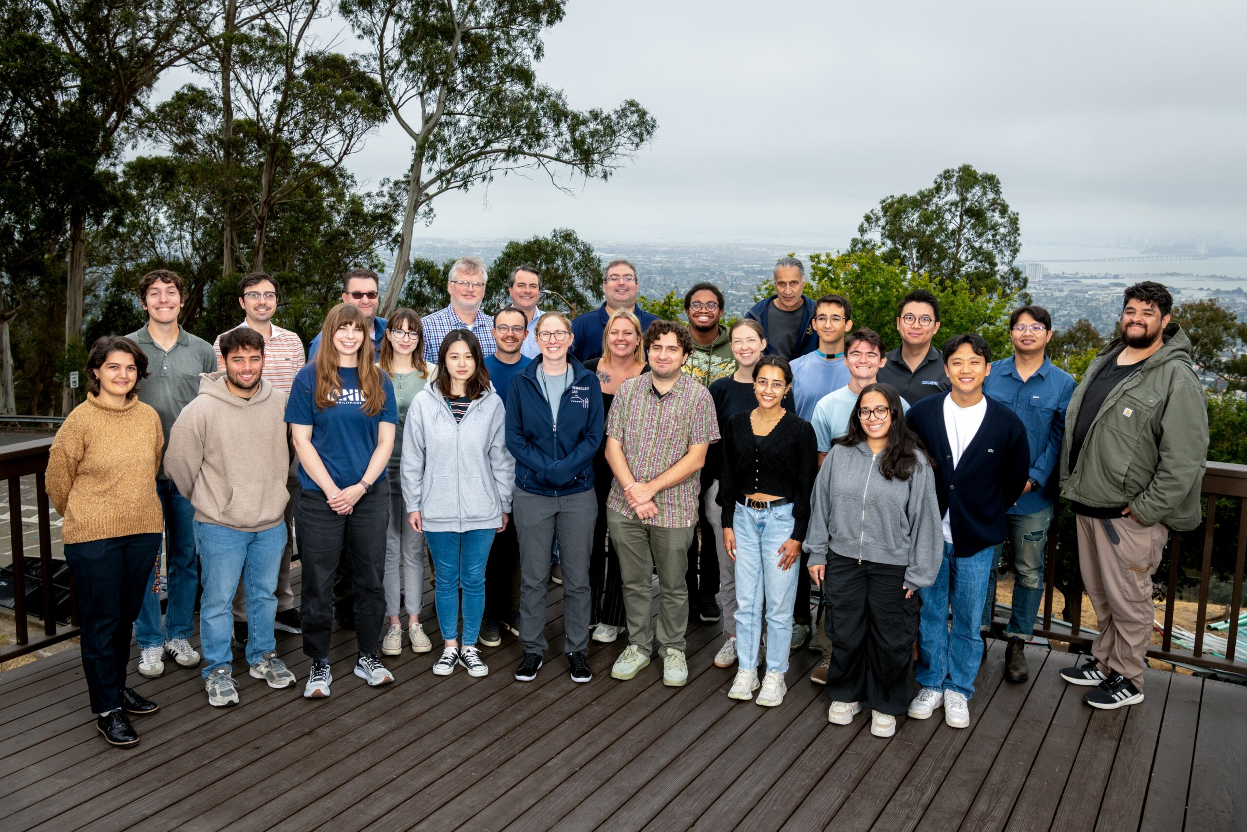
Hands-on workshop to extend the impact of SYNAPS-I
“Synchrotron user facilities are stewards of quite possibly the most scientific data in the world,” said David Abramov, a research software engineer with the ALS Computing group.
Indeed, as a microtomography beamline scientist, Clark might support one user’s experiments examining spacecraft heat shields, then another’s on dozens of heads of lettuce. Other typical samples include battery cathodes, pistachios, and jumping spiders. The scores of samples add up to a deluge of data.
“Some microtomography power users can collect 3,000 scans in a five-day period of beamtime, which is up to 150 TB of raw data alone—not including any processed results,” Abramov added.
The power user in question, Eli Hiss, is a mechanical engineer/materials scientist at NASA Ames studying heat shielding for spacecraft such as the recent Artemis missions. He isn’t the only user generating astounding quantities of data. As beamline scientists, controls, and computing staff work together to support even faster and higher resolution detectors, the amount of data is poised to explode. The next challenge is figuring out how to process the data much more efficiently, especially during beamtime when users can steer experiments based on real-time feedback.
Other users at the workshop study energy, life, earth, and materials sciences. Their research differs, but they face common bottlenecks in data processing. Together with beamline scientists and software engineers, they are improving the tools to ease those bottlenecks.
Users enthusiastic to improve the workflow
During the workshop, Abramov unveiled an open-source platform, an interface for users to access and share feedback on the computer vision tools. Researchers can use the software for segmentation by hand as well as training custom AI segmentation models. These types of tools typically cost up to $10,000 to load on an individual user’s computer. Now, people like Hiss, along with anyone generating tomographic imaging data, can access the resources for free.
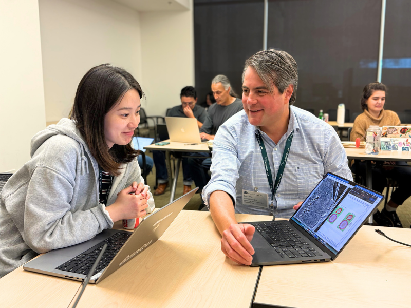
The workshop attendees contributed valuable feedback on what tools and features to add next into the software. Even after segmentation, there are more analysis steps and measurements users perform, which they provided insight on. The more developers listen to the users, the better they can align the development process to user needs, and generate a better experience for visiting researchers across scientific disciplines.
When testing out the new software, a few users remarked that they love being able to easily undo their labeling without disrupting their workflow, unlike other tools they have used. Also, the interface was “very intuitive,” with one user complimenting the “clean sidepanel,” as well as the polygon tool, option to grow the shape, and ability to change the class of a shape.
Overall the responses were positive, with the energy science users noting, “The software is great for speeding up the annotation process, is more user friendly, and we are definitely interested in continuing to use it.”
Users also commented about new features that were demonstrated by Alex Hexemer and fellow team members, Computer Staff Scientist Petrus Zwart and ALS Doctoral Fellow Pradyumna Elvarthi, saying they were excited to try out labeling a “small subset” of the image really quickly before applying the process to the full image and iterating.
“This seems like it will help the task go quicker,” the user said.
Users also provided a variety of feedback about what improvements they would like to see developed further, and are excited to have the tool integrated with the tomography workflow. UC Berkeley graduate student and user Sophie Hanson remarked that she wants to reach out to other researchers who have annotated arachnids, insects, and arthropods for their data to help train a model they can all benefit from at their next beamtime.
As Clark joked, “Now you don’t have to watch a sitcom about a group of friends. You’ll have time to have friends!” And, these friends are excited to tackle the next challenges in energy sciences, earth sciences, life sciences, and materials sciences.