by Brooke Kuei
Many important chemical reactions, such as those in batteries and catalysts, occur at interfaces where a solid material meets a liquid. However, these regions are notoriously difficult to study because the thin interface is buried beneath a thick layer of liquid, making it challenging to detect signals from the interface amidst the signal of its surroundings without a high dose of damaging photons or electrons.
As reported in a recent Nature Communications study, Berkeley Lab researchers developed a new x-ray technique based at the Advanced Light Source (ALS) called pattern-enhanced resonant soft x-ray scattering (PE-RSoXS). The novel method dramatically enhances weak signals from buried interfaces by engineering nanoscale patterns directly into the sample. The Center for High Precision Patterning Science (CHiPPS), an Energy Frontier Research Center (EFRC) funded by the U.S. Department of Energy (DOE), supported this work in collaboration with various Berkeley Lab programs and focuses on developing fundamental science for high-precision patterning technologies used in next-generation microelectronics and advanced manufacturing.
“The concept we demonstrated is what we call sample-as-optics,” said Cheng Wang, staff scientist at ALS Beamline 11.0.1.2 and lead of the characterization thrust for the CHiPPS EFRC. “Traditionally, the optics and the sample are separate. Here, we intentionally design nanostructures in the sample so the interaction between the x-rays and the material is amplified by the optical properties of that structure.”
To demonstrate the approach, the researchers collaborated with the Molecular Foundry and Center for X-Ray Optics (CXRO) to fabricate nickel electrodes patterned with an array of nanoscale structures, choosing nickel because it is both stable during lithography and a well-studied electrochemical model system. Using resonant soft x-ray scattering (RSoXS) at Beamline 11.0.1.2, the team studied the oxygen evolution reaction at the electrode–electrolyte interface, demonstrating that the periodic nanostructures produced coherent scattering—similar to the diffraction from repeating crystal lattices in protein crystallography—causing the diffraction intensity to scale with the square of the number of repeating units.
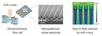
In the patterned region used in this experiment, coherent scattering produced signal amplification approaching five orders of magnitude, enabling detection of subtle chemical and structural changes at the buried interface that were supported by simulations performed at the National Energy Research Scientific Computing Center (NERSC). The measurements are also fast, and the radiation dose is spread out over a large number of structures, which means the average dose is significantly reduced compared to microscopic methods.
This creative approach to advancing synchrotron science highlights the collaborative, team science model at Berkeley Lab and opens new opportunities for studying a wide range of materials systems. “This method isn’t limited to electrochemistry,” Wang said. “Any time you can engineer a patterned structure, whether for catalysis, semiconductor materials, or even biological systems, you can potentially use it to enhance the signal and study processes that were previously too small or too buried to observe.” Ongoing efforts within the Liquid Sunlight Alliance (LiSA), for example, are exploring electrochemical systems that can take advantage of this approach.
H. Li, K. Andrle, Q. Zhang, I. A. Cordova, Y. Yang, Z. Peng, F. Yang, G. Freychet, S. Dhuey, A. Hexemer, B. A. Helms, W. Chao, B. La Fontaine, R. Ruiz, J. Guo, W. Yang, J. Yano, and C. Wang, “Pattern-enhanced Resonant Soft X-ray Scattering for Operando Monitoring of Electrochemical Solid–Liquid Interfaces,” Nature Communications (2026), doi:10.1038/s41467-026-69852-9.