In 1994, a virus emerged in Hendra, Australia, causing respiratory and neurological diseases. It was transmissible from horses to humans, with a mortality rate of 57% in humans and 89% in horses. In 1999, a similar virus, transmitted through domesticated pigs, caused over 100 human deaths in Sungai Nipah, a Malaysian village. The Hendra and Nipah viruses are classified as deadly members of the Paramyxoviridae family, under the emerging genus Henipavirus. This genus was recently expanded to include a third species (Cedar virus), and evidence exists for 19 additional species in Africa.
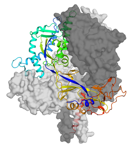
Paramyxovirus infection of host cells is mediated by the fusion protein, F, which is embedded in the viral particle membrane. The bulk of the F protein, the ectodomain, protrudes from the membrane’s surface and undergoes a dramatic refolding to merge the virus and host membranes. At ALS Beamline 8.2.2, researchers used macromolecular crystallography to study the structure of the Hendra F protein ectodomain in its prefusion form.
The resulting atomic-level model allowed the researchers to visualize the F protein’s shape and determine its surface chemical properties. They also found that the overall structure of the Hendra F protein is very similar to that of another, relatively distant member of the Paramyxoviridae family, parainfluenza virus V, with two small regions of difference, providing valuable insight into the mechanistic steps in the virus entry process. With this information, the researchers were able to engineer a stabilized form of the Hendra F protein that more closely mimics the shape of the molecule on the virus surface, which should make it more potent at eliciting neutralizing antibodies as part of a vaccine.

Work performed at ALS Beamline 8.2.2 (Berkeley Center for Structural Biology).
J.J.W. Wong, R.G. Paterson, R.A. Lamb, and T.S. Jardetzky, “Structure and stabilization of the Hendra virus F glycoprotein in its prefusion form,” PNAS 113, 1056 (2016).