Researchers found that a symbiont capable of fixing nitrogen (turning it into a biologically usable form) has evolved into an organelle—an intrinsic part of the algae cells that host it. The discovery is of great interest for understanding organelle genesis and for efforts to engineer agricultural plants with built-in nitrogen-fixing capabilities. Read more »![]()
![]()
Scientists Discover First Nitrogen-Fixing Organelle
In two recent papers, an international team of scientists describe the first known nitrogen-fixing organelle within a eukaryotic cell. The organelle is the fourth example in history of primary endosymbiosis—the process by which a prokaryotic cell is engulfed by a eukaryotic cell and evolves beyond symbiosis into an organelle. Read more »
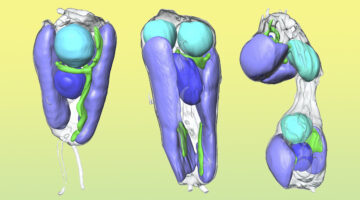
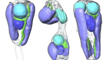
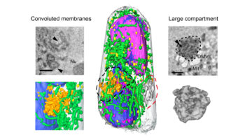
Nitrogen-fixing organelle in a marine alga

A nitrogen-fixing organelle, or “nitroplast,” has been identified in a marine alga on the basis of intracellular imaging and proteomic evidence. This discovery sheds light on the evolutionary transition from endosymbiont to organelle. The image depicts the cell architecture and synchronized cell division of the alga Braarudosphaera bigelowii with nitroplast UCYN-A (large brown spheres). Read more »
A New Pathway for Clearing Misfolded Proteins
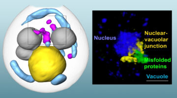
Researchers integrated several approaches, such as cryogenic 3D imaging at the ALS, to define a novel cellular pathway—involving a shared “garbage dump”—for clearing misfolded proteins from cells. The pathway is a potential therapy target for age-related diseases like Alzheimer’s, Huntington’s, and Parkinson’s diseases. Read more »![]()
![]()
Safely Studying Dangerous Infections Just Got a Lot Easier
Researchers have cranked up the speed of imaging infected cells using soft x-ray tomography, a technique that can generate incredibly detailed, three-dimensional scans. The approach gives an easy way to quickly examine how cells’ internal machinery responds to SARS-CoV-2, or other pathogens, as well as how the cells respond to drugs designed to treat the infection. Read more »
Rapid 3D Visualization of Lung Cells Altered by SARS-CoV-2
In this work, researchers illustrated the potential of soft x-ray tomography to rapidly characterize and quantify the structural changes induced in cells infected by SARS-CoV-2, revealing profound alterations of the subcellular architecture induced by viral infection over time. Read more »
3D Whole-Cell Mapping of Insulin Secretion
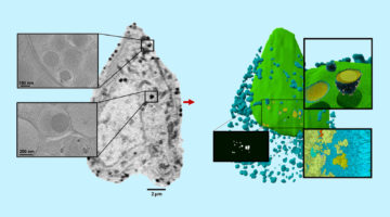
Researchers used soft x-ray tomography to gain a 3D whole-cell view of how insulin-producing pancreatic cells react upon exposure to glucose and a diabetes drug. The approach enables direct quantification of intracellular responses before, during, and after cell stimulation, providing new insights into how drugs alter cell function. Read more »![]()
![]()
Unique X-Ray Microscope Reveals Dazzling 3D Cell Images
Researchers used soft x-ray tomography to reveal never-before-seen details about insulin secretion in pancreatic cells taken from rats. By quantifying subcellular rearrangements in response to drugs, the results are an important first step for bridging the longstanding gap between structural biology and physiology. Read more »
How Proteins Remodel DNA in Bacteria under Stress
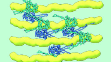
Multiscale, multimodal visualization techniques at the ALS enabled researchers to clarify how proteins remodel bacterial DNA in response to stressful environments. The discovery could lead to new strategies for controlling microbial behavior and, eventually, new ways to fight bacterial infections. Read more »![]()
![]()
Study Gains New Insight Into Bacterial DNA Packing
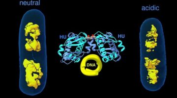
When bacteria are put in different environments, their genes start to adapt remarkably quickly because the proteins making up their chromosomes can pack and unpack rapidly. Researchers have now imaged this process at the molecular level, a discovery that could eventually enable scientists to develop strategies to control microbial behavior. Read more »